

Blue Quartz
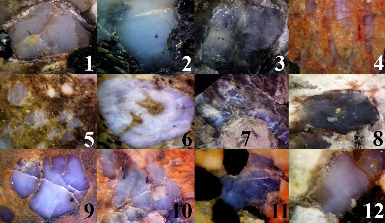
Polished sections of various blue quartz occurrences as seen under the stereographic microscope: 1-3 Albești metagranite (Romania); 4 Cosăuți granite (Moldova); 5-6 Pietrosu Bistriței porphyroid (Romania); 7 Rio dos Remédios alkali rhyolite (Bahia, Brazil); 8 Lalu granite (Romania); 9 Llano rhyolite (Texas, USA); 10 Milbank granite (South Dakota, USA); 11 Småland granite (Sweden); 12 Valea Bolovanului granite (Romania).
Quartz is the most familiar mineral on Earth: colorless, glassy, and seemingly ordinary, it forms the backbone of granites, gneisses, sandstones, and countless veins. Yet quartz is also a quiet chronicler. Its subtle color variants — smoky, rose, amethyst, citrine — each encode a different story about the chemistry and physics of the environment in which the crystal grew. Among these variants, few are as visually arresting or as scientifically puzzling as blue quartz.
Blue quartz is exactly what its name suggests: a coarsely crystalline (macrocrystalline) variety of quartz that appears blue. The hues range widely — from a dilute, almost ethereal aquamarine, through milky "dove-blue" and smoky blue-grey, to an intense, sapphire-like violet-blue (Szopa et al., 2017; Li et al., 2020; Pantia et al., 2022). Very often the color is accompanied by a soft, cloudy, glowing quality known as opalescence, a milky sheen that seems to hover within the crystal (Emerson, 1899; Nockolds, 1931; Landenberger, 1996; Pantia et al., 2022). To many observers the two properties — the blueness and the opalescence — feel inseparable, and as we shall see, that intuition turns out to be physically correct.
What makes blue quartz genuinely unusual is that its color is not a chemical property in the ordinary sense. Most colored minerals owe their hues to absorption: certain ions or defects in the crystal soak up particular wavelengths of light, and we see what is left over. Blue quartz is different. Its color comes primarily from the scattering of light — the same physical process that paints the daytime sky blue — by a dense swarm of vanishingly small particles trapped inside the crystal (Iddings, 1904; Jayaraman, 1939; Zolensky et al., 1988; Seifert et al., 2011). This is the single most important idea in the whole subject, and the rest just explores its consequences.
Because the color is a physical effect rather than a chemical one, it is useful to define blue quartz by that physical criterion. A modern, mechanism-based definition runs as follows: blue quartz is a macrocrystalline variety of SiO₂ whose blue color results from the scattering of short-wavelength light by submicron to nanometric inclusions, structural discontinuities, or lattice defects, regardless of its geological age, host rock, or mode of origin. This definition does two important things. First, it deliberately excludes quartz that looks blue because it contains blue mineral inclusions — such as dumortierite, tourmaline, or magnesio-riebeckite — since those minerals color the quartz by absorption, a fundamentally different mechanism. Second, it excludes the fine-grained and non-crystalline forms of silica, chalcedony and opal, which belong to a different structural and geological family altogether (their microstructures and low-temperature, often watery origins set them apart from the high-temperature, rock-forming quartz we are concerned with here).
Blue quartz has fascinated geologists for well over a century. One of the classic early descriptions came from J. P. Iddings in 1904, working on the striking "llanite" porphyry of Llano County, Texas, whose blue quartz phenocrysts remain a textbook example to this day (Iddings, 1904; Zolensky et al., 1988). Half a world away, in the charnockites and gneisses of southern India, N. Jayaraman puzzled over the same phenomenon in 1939, and his careful observations on the color’s behavior still inform the debate (Jayaraman, 1939). Perhaps the most poetic acknowledgement of blue quartz is embedded in a place-name: across the Iberian Peninsula, a vast belt of Cambro-Ordovician gneisses and volcanic rocks is called the Ollo de Sapo — Spanish for "toad's eye" — after the large, staring, deep-blue quartz megacrysts that speckle the rock (Díez Montes et al., 2010; von Raumer & Stampfli, 2018).
Far from being a mere curiosity, blue quartz carries real geological information. Because it tends to form under specific conditions — high temperatures, titanium-rich melts or fluids, and comparatively "dry" (water-poor) environments — its presence can act as a field-visible signpost to the deep, hot, and often ancient parts of the continental crust (Seifert et al., 2011; Raumer & Stampfli, 2018). Geologists have used it as a petrogenetic indicator (a clue to how a rock formed), as a tectonic marker (a clue to the large-scale setting), and even as an exploration guide near certain uranium and gold deposits (Zolensky et al., 1988; Hillacre, 2018; Choquette, 2021). It has been reported on every continent and across nearly three billion years of Earth history — from Archean greenstone belts to Pleistocene rhyolites — which makes it not just beautiful, but a genuinely global and deep-time phenomenon (Samsonov et al., 1993; Rossotti et al., 2002; Seifert et al., 2011).
The sections that follow tell the story in stages. We begin with the physics of the color itself — why blue, and why cloudy. We then turn to the central and still-unresolved scientific controversy: exactly how big must the trapped particles be to produce the effect? Next we identify the "suspects" — the tiny mineral inclusions responsible for the scattering — and ask where they come from. Finally, we zoom out to the planetary scale and map where, and when, blue quartz appears in the geological record.
The Physics of the Blue: Unraveling the Scattering Mechanism
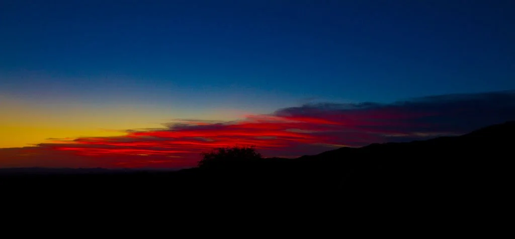
A beautiful example of atmospheric Rayleigh scattering during sunrise/sunset (Photo: https://beautifulnow.is/discover/nature-science/science-explains-why-red-skies-happen-at-sunrise-and-sunset-and-why-they-are-all-more-beautiful-in-autumn).
To understand blue quartz, it helps to start with a familiar question: why is the sky blue? The answer, worked out in the nineteenth century, is that molecules and tiny particles in the atmosphere scatter sunlight, and they scatter short (blue) wavelengths far more strongly than long (red) ones. Blue light is flung in all directions across the sky, so wherever we look — away from the Sun — we see blue. Blue quartz is, in a very real sense, a piece of sky trapped in stone. The same physics that colors the atmosphere colors the crystal (Nassau, 1983; Zolensky et al., 1988).
Scattering vs. absorption
The first and most important distinction is between scattering and absorption, the two great families of color-producing mechanisms.
In absorption, a material removes certain wavelengths from white light by converting their energy into other forms (usually heat or electronic transitions). The colors of amethyst (iron-related color centers), of emerald (chromium), or of most blue minerals arise this way. What reaches your eye is the leftover, un-absorbed light.
In scattering, no light is absorbed. Instead, incoming light strikes a small obstacle — a particle, a bubble, a density fluctuation — whose electrons are set oscillating by the light's electromagnetic field. These oscillating electrons act as tiny antennas, re-radiating the light in new directions but at the same wavelength (Tilley, 2011). If the obstacles are very small compared with the wavelength of light, they re-radiate blue much more efficiently than red, and the material takes on a blue appearance in reflected or side-scattered light. This is the mechanism at work in blue quartz.
The consensus that scattering — not absorption — is responsible for the color of rock-forming blue quartz is broad and long-standing, supported by observations spanning more than a century (Iddings, 1904; Jayaraman, 1939; Kennard & Howell, 1941; Zolensky et al., 1988; Seifert et al., 2011).
The three scattering regimes
Physicists classify scattering by the size of the scattering centers relative to the wavelength of visible light (roughly 400–700 nanometers, where a nanometer is a billionth of a meter).
Rayleigh scattering occurs when the particles are much smaller than the wavelength — conventionally less than about one-tenth of it, or below roughly 50–70 nm. In this regime the scattered intensity is proportional to λ⁻⁴ (one over the wavelength to the fourth power). That steep dependence is the key: blue light (short λ) is scattered far more strongly than red light (long λ), which is precisely why both the sky and blue quartz are blue. A dense population of such sub-wavelength particles can impart a distinct blue tint to an otherwise transparent host (Nassau, 1983; Zolensky et al., 1988).
Tyndall scattering is closely related but is usually invoked for somewhat larger particles, roughly in the 40–900 nm range. The wavelength dependence (λ⁻⁴) is preserved, so blue is still favored, but because the particles present a larger cross-section, the overall scattering is more intense. This regime tends to produce the pronounced milky, opalescent appearance so characteristic of blue quartz (Nassau, 1983; Seifert et al., 2011).
Mie scattering takes over when the particles become comparable to, or larger than, the wavelength of light. Here the physics becomes more complicated: the wavelength preference weakens, and scattering becomes more "neutral" and directional, producing whitish or milky tones rather than a clean blue. In blue quartz, Mie-type scattering is generally associated with the larger inclusions that produce bright flashes or shimmering bands (chatoyancy), while the true blue color remains the work of the finer Rayleigh–Tyndall population (Nassau, 1983).
Why the color depends on the inclusions' identity
Size is not the only thing that matters. A particle scatters strongly only if its refractive index differs substantially from that of the surrounding host — a mismatch expressed as Δn. As a rule of thumb, meaningful scattering requires Δn of at least about 0.1. (For comparison: air bubbles in water, with Δn ≈ 0.33, scatter strongly and make foam look white; certain polymer nanoparticles in glass, with Δn ≈ 0.05, scatter almost imperceptibly.)
This is why rutile (titanium dioxide, TiO₂) is repeatedly singled out among the possible inclusions. Rutile has an exceptionally high refractive index — around 2.6–2.9, one of the highest of any common rock-forming mineral — whereas quartz sits near 1.55. The resulting contrast, Δn ≈ 1.1–1.4, is enormous, far more than enough to make even sparse, tiny rutile particles powerful scattering centers (Seifert et al., 2011). A crystal can therefore look intensely blue with only a modest quantity of rutile, provided the particles are the right size and densely distributed.
The fingerprints of a scattering origin
How can a geologist confirm, in practice, that a blue quartz owes its color to scattering rather than to absorption or some other cause? Two simple, powerful tests emerge directly from the physics.
The transmitted–reflected color test. Because short wavelengths are preferentially scattered out of the transmitted beam, light that passes through blue quartz has had its blue component depleted, and so emerges looking warm — reddish, yellowish, or brownish. The very same grain, viewed in reflected or side-scattered light, looks blue. This blue-in-reflection, warm-in-transmission mismatch is a hallmark of Rayleigh- and Tyndall-type scattering, and it has been documented in occurrence after occurrence around the world (Kennard & Howell, 1941; Jayaraman, 1939; Barker & Burmester, 1970; Zolensky et al., 1988; Pantia & Filiuță, 2021; Pantia et al., 2022). It is exactly analogous to the sky (blue, scattered) and the setting Sun (red, transmitted through a long path of atmosphere).
The dark-background test. Scattered blue is most vivid against a dark, non-reflective backdrop. The blue sky is brilliant against the black of space; blue eyes appear blue because a dark layer behind the iris absorbs the un-scattered light; and blue quartz looks its bluest when it sits on or against a dark matrix, where bright background reflections do not wash out the delicate scattered signal (Nassau, 1983). This is why the same quartz grain can look strikingly blue in a dark, biotite-rich rock and disappointingly pale in a light-colored one.
Opalescence: the color’s constant companion
The milky opalescence that so often accompanies blue quartz is not a separate phenomenon — it is the same phenomenon seen slightly differently. The dense population of submicron scattering centers that generates the blue color also scatters light diffusely throughout the grain, producing the soft, glowing, cloudy texture. This is why opalescence and blue color are so tightly linked: a grain may be opalescent without being obviously blue, but a genuinely (scattering-)blue grain is essentially always opalescent (Moore, 1911; Pantia & Filiuță, 2021; Pantia et al., 2022). The coincidence of the two is itself a useful diagnostic that the color is truly scattering-based.
A minor alternative: structural color
For completeness, a small number of blue quartz occurrences may owe their color to structural color — thin-film interference, in which light reflecting off the two surfaces of an ultra-thin film (for example, a carbonaceous coating on a grain) interferes constructively for blue wavelengths (Bjørlykke, 1983). This mechanism is rare, because it demands very uniform, nanometer-scale films that geological processes rarely produce or preserve. When it does occur, it usually announces itself through iridescence — a metallic-looking color that shifts with viewing angle — rather than the diffuse, milky blue of true scattering. It is a real but minor contributor to the global blue-quartz spectrum.
The Core Controversy: Does Size Matter?
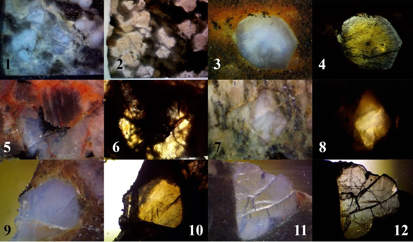
Color differences in reflected/transmitted light: 1-2 Albești granite (Romania); 3-4 Llano rhyolite (Texas, USA); 5-6 Milbank granite (South Dakota, USA); 7-8 Pietrosu Bistritei porphyroid (Romania); 9-10 and 11-12 Rio dos Remédios alkali rhyolite (Bahia, Brazil).
Everyone agrees that blue quartz is colored by scattering. But agreement dissolves the moment one asks a deceptively simple question: exactly how big are the particles doing the scattering? This is the central, still-unresolved controversy of the subject, and it turns out to matter a great deal — because the size of the scattering centers determines which scattering regime dominates, and therefore whether the tidy "Rayleigh scattering" explanation actually holds up.
The theoretical expectation
As we saw, pure Rayleigh scattering — the classic explanation for a clean blue — requires particles much smaller than the wavelength of light. The textbook threshold is about one-tenth of the wavelength, meaning below roughly 50–70 nm for visible light. Some authors tighten this considerably, to one-twentieth of the wavelength, or below about 27–28 nm (Seifert et al., 2011). Others are more generous, allowing particles up to around 300 nm to still count within a broadened Rayleigh–Tyndall range, depending on the refractive-index contrast and the shape of the particles (Nassau, 1983).
So there is already a spread in the "allowed" sizes even before we look at real rocks. When we do, the picture becomes more complicated still.
What the measurements actually show
More than a century of observations has produced a remarkably scattered set of size estimates for the inclusions in blue quartz:
- Iddings (1904) cited inclusions smaller than about 800 nm.
- Moore (1911) referred to inclusions smaller than half a wavelength of light.
- Jayaraman (1939) proposed 200–500 nm as the size range for maximum scattering.
- Wise (1981) described ilmenite lamellae only 0.02–1 μm (20–1000 nm) thick, but up to hundreds of micrometres long.
- Zolensky et al. (1988) reported ilmenite inclusions averaging about 60 nm, at astonishing densities of up to 125 particles per cubic micrometer.
- Herz & Force (1987) described ilmenite plates up to about 250 nm.
- Seifert et al. (2011), using modern high-resolution techniques, found mica flakes under 100 nm thick, ilmenite of 50–100 nm, and rutile as fine as 25–50 nm.
The problem is immediately apparent. If one applies the strict Rayleigh threshold (below ~50 or even ~27 nm), then many of these reported inclusions are simply too large to produce a blue color by pure Rayleigh scattering. A 250 nm ilmenite plate, taken at face value, ought to scatter light almost neutrally (Mie-style) and make the quartz look white or grey, not blue. So how can such "oversized" inclusions be reconciled with the vivid blue that is actually observed? Several resolutions have been proposed, and together they dissolve the apparent paradox.
Resolution 1: it's the smallest dimension that counts
Many of the offending inclusions are not equant blobs but thin plates or slender needles. For scattering, the physically relevant dimension of an elongate or platy particle is its smallest one — the thickness of the plate or the diameter of the needle, not its length. A 250 nm ilmenite plate that is only 50 nm thick can behave as a Rayleigh scatterer along its thin axis, even though it looks "large" in a two-dimensional image (Wise, 1981). Much of the apparent size problem evaporates once shape is taken into account.
Resolution 2: the power of refractive-index contrast
As noted earlier, a large refractive-index mismatch enhances scattering and effectively pushes the usable size limit upward. Because rutile's contrast with quartz is so extreme (Δn ≈ 1.1–1.4), rutile particles somewhat larger than the classic Rayleigh threshold can still scatter blue light efficiently. The strict size limits derived for low-contrast systems simply do not apply with full force to high-contrast inclusions like rutile and ilmenite (Seifert et al., 2011).
Resolution 3: Tyndall scattering widens the window
The Tyndall regime explicitly accommodates larger particles — up to around 900 nm — while preserving the crucial λ⁻⁴ preference for blue (Nassau, 1983). Once Tyndall scattering is admitted alongside Rayleigh scattering, the "acceptable" size window broadens dramatically, and most of the reported inclusion sizes fall comfortably inside it. In reality, blue quartz almost certainly reflects a combination of Rayleigh and Tyndall scattering rather than either one alone.
Resolution 4: density, not just size
The intensity of the color depends not only on how big the individual particles are, but on how many of them there are per unit volume. A dense cloud of small scatterers can produce a strong blue even if a minority of the particles stray toward the upper size limit (Moore, 1911; Kennard & Howell, 1941; Herz & Force, 1987). The extraordinary densities reported by Zolensky et al. (1988) — over a hundred particles per cubic micrometer — illustrate just how crowded these scattering populations can be.
Resolution 5: the crystal itself can scatter
Finally, discrete mineral inclusions may not be the whole story. Density fluctuations — subtle, sub-wavelength variations in composition or structure within the quartz lattice itself — can act as scattering centers even where no separate mineral phase is present (Kerker, 1969; Gopal, 2000). From this broader viewpoint, the "size" of any individual inclusion may matter less than the overall, integrated field of nanoscale heterogeneity pervading the crystal.
The state of the debate
Taken together, these considerations show that the size controversy is not a fatal flaw in the scattering model, but a reminder of its subtlety. Blue quartz color is not produced by a single, idealized particle size, but by the combined effect of a whole population of scatterers — spanning a range of sizes, shapes, densities, and optical contrasts — operating across the Rayleigh and Tyndall regimes at once. What remains genuinely open, and what modern instruments (electron and transmission electron microscopy, in particular) are now beginning to resolve, is the precise size distribution and density of these particles in different occurrences, and how those parameters vary with geological setting. Until such systematic, quantitative characterization is done across many localities, the exact recipe that turns a given quartz crystal blue will remain one of the field's most intriguing loose ends.
The Suspects and Their Origins: Unmasking the Scattering Centers
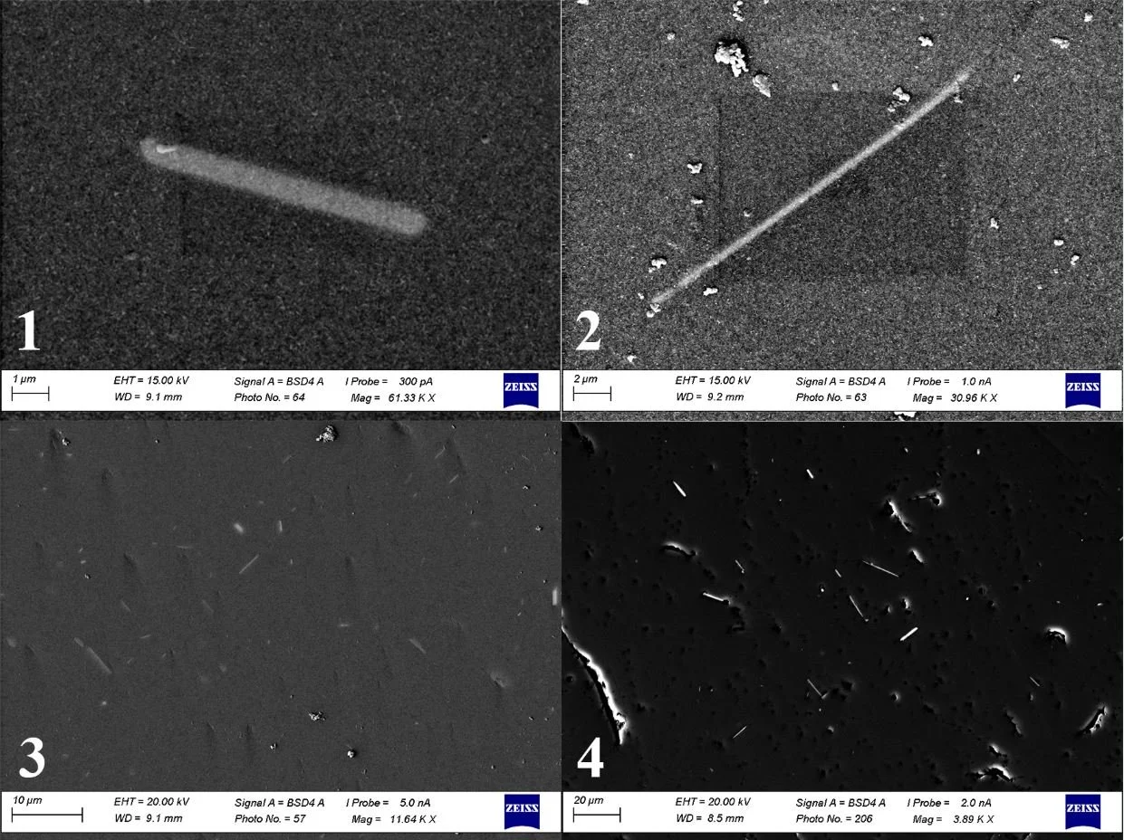
Submicron rutile inclusions in: 1 Albești granite (Romania), 8/0.63 μm; 2 Llano rhyolite (Texas, USA) 21/0.37 μm; 3 Milbank granite (South Dakota, USA) 3.78/0.07 μm on average; 4 Pietrosu Bistriței porphyroid (Romania) 8.5/0.33 μm on average.
If scattering produces the color, then something must be doing the scattering. Identifying these "suspects" — the tiny inclusions embedded in the quartz — and working out where they came from is one of the richest and most contested parts of the blue-quartz story. It is also a story with a cautionary subplot about how scientific ideas can harden into dogma.
The prime suspect: rutile
By a wide margin, the inclusion most often blamed for blue quartz is rutile (TiO₂), typically in the form of submicron needles. Rutile has been named in descriptions dating back to Emerson (1899) and Watson & Taber (1913) and continuing through the modern literature (Seifert et al., 2011; Hillacre, 2018). It is such a favorite that "rutilated blue quartz" has become almost a stock phrase, and rutile's exceptionally high refractive index (Section 2) gives it a genuine theoretical claim to the role.
But there is a catch. Much of rutile's apparent dominance may reflect citation habit rather than direct evidence. Many papers that attribute blue color to rutile do so by echoing earlier studies, without independently checking the mineralogy in their own samples. A telling example: Gentle (1977) inferred that rutile caused the blue color in certain South African granitoids largely by analogy with Howie's (1955) work on charnockites — a different rock type entirely — rather than by direct observation. Once such an attribution enters the literature, it tends to propagate, and rutile's reputation may be partly self-reinforcing.
A broader line-up
When researchers actually look, the roster of scattering inclusions turns out to be far more diverse than the rutile-centric narrative suggests.
Ilmenite (FeTiO₃) is a major alternative, documented as a key — sometimes the dominant — scattering phase by many workers (Ross, 1941; Wise, 1981; Herz & Force, 1987; Zolensky et al., 1988; Seifert et al., 2011; Pantia & Filiuță, 2021). At the famous Llano rhyolite, ilmenite, not rutile, is the principal culprit (Zolensky et al., 1988).
Micas (biotite, muscovite) have been reported as nano-scale scatterers, and Seifert et al. (2011) made the striking observation that mica is actually the most numerous submicron inclusion in some blue quartz samples — even though the color is still usually credited to rutile or ilmenite, because scattering theory so strongly favors high-refractive-index minerals over micas.
Beyond these, tourmaline, zircon, and apatite have been invoked in various granitic and metamorphic settings (Parker, 1962; Barker & Burmester, 1970; Ross, 1941; Zolensky et al., 1988), and graphite (Seifert et al., 2011; O'Brien et al., 2015) and fluid inclusions (Allison, 1925; Nockolds, 1931; Andersen et al., 1990) round out the cast. The honest conclusion is that no single mineral is the cause of blue quartz; rather, a family of submicron phases can each play the role, with rutile and ilmenite favored on theoretical grounds but by no means exclusive.
Where do the inclusions come from?
Identifying the suspects is only half the investigation. The deeper question is how these tiny particles came to be trapped inside the quartz in the first place. Several distinct origins have been proposed, and many blue quartz crystals probably record more than one.
Exsolution. The most frequently cited mechanism is exsolution. Quartz crystallizing at high temperature can take up small amounts of titanium into its lattice; as the crystal later cools, that titanium becomes insoluble and precipitates out as a fine dusting of nanoscale rutile needles (Watson & Taber, 1913; Parker, 1962; Herz & Force, 1987; Seifert et al., 2011). This elegant process neatly ties blue quartz to titanium-rich, high-temperature systems, and it is a genuine and important pathway for rutile.
But not for ilmenite — a cautionary correction. Here the literature contains a persistent error worth highlighting. One of the most cited papers on blue quartz, Zolensky et al. (1988), attributed the light-scattering ilmenite of the Llano rhyolite to exsolution from the host quartz. This cannot be correct. Ilmenite has a compact hexagonal structure in which iron and titanium occupy octahedral sites in equal proportion — a structure fundamentally incompatible with the quartz framework (Wenk & Bulakh, 2016). Moreover, quartz is essentially pure SiO₂, carrying only trace titanium and aluminum; it simply never contains enough iron and titanium to nucleate ilmenite, and even if it did, ilmenite requires a coupled mobility of iron and titanium oxides that quartz cannot supply (Lindsley, 1991). Ilmenite inclusions in blue quartz must therefore be trapped or precipitated by some other process, not exsolved from the lattice — an important correction to a widely repeated claim.
Strain and deformation. Deformation can also generate scattering centers. Squeezing and shearing a crystal can nucleate and align new inclusions, or create microfractures, deformation bands, and dislocation-rich zones that scatter light in their own right (Crickmay, 1936; Castro de Machuca & López, 2017). Experimental work by Thomas & Nachlas (2020) showed that "dry" recrystallization of titanium-rich quartz, driven by migrating grain boundaries, precipitates randomly oriented rutile needles in the wake of the moving boundaries — suggesting that irregular, acicular rutile can be a signature of high-temperature, water-poor recrystallization rather than simple magmatic growth.
Syngenetic entrapment. In other cases, the inclusions are simply pre-existing microcrystals — of rutile, ilmenite, or other phases — that were already floating in the melt and became trapped inside the quartz as it grew around them (von Vultée, 1955; Seifert et al., 2011). A related idea invokes crystallization at the quartz–melt boundary, where local build-ups of elements like magnesium and iron form phases that the quartz cannot incorporate and so entombs as inclusions (Seifert et al., 2011; Thomas & Nachlas, 2020). This is especially relevant to the peraluminous, sediment-derived magmas that host blue quartz in belts like the Ollo de Sapo, where inherited titanium minerals from the source rocks can be swept up by rapidly growing quartz (Díez Montes et al., 2010; von Raumer & Stampfli, 2018).
The common thread: heat, titanium, and speed
Different as these mechanisms are, they converge on a shared set of conditions. Blue quartz overwhelmingly forms where temperatures are high (commonly 700–900 °C, as gauged by titanium-in-quartz thermometry), where titanium is abundant, and where crystallization or recrystallization is rapid enough to trap the scattering particles before they can grow too coarse and lose their optical punch (Wark & Watson, 2006; Seifert et al., 2011; Thomas & Nachlas, 2020). Whatever the exact suspect and whatever its precise origin, the outcome is the same: millions of sub-wavelength particles, densely packed within a single crystal, collectively scattering short-wavelength light and turning ordinary quartz an extraordinary blue.
A Global Puzzle: Mapping the Occurrences of Blue Quartz
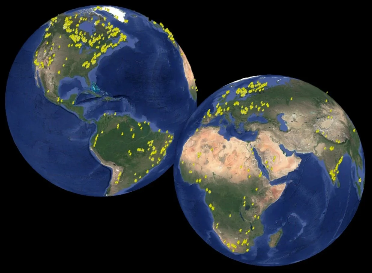
The global distribution of magmatic and metamorphic blue quartz occurrences.
Blue quartz is not a local oddity confined to one lucky outcrop. It has been reported from every continent, in rocks ranging in age from the Archean to the Pleistocene, and across an astonishing variety of geological settings (Samsonov et al., 1993; Rossotti et al., 2002; Seifert et al., 2011). Yet its distribution is anything but random. When the scattered reports are gathered and mapped, blue quartz reveals striking patterns in where and when it forms — patterns that turn a beautiful mineral into a genuine tool for reading the history of the continents.
A strongly uneven distribution
Although blue quartz occurs worldwide, it clusters conspicuously in certain kinds of terrain. Four settings recur again and again:
- Proterozoic anorogenic magmatic provinces — belts of "A-type" and rapakivi granites and massif-type anorthosites, emplaced far from active plate collisions, in hot, dry, extensional or transtensional settings (Zolensky et al., 1988).
- Granulite-facies metamorphic terranes — the deepest, hottest, driest levels of ancient mountain belts, where blue quartz can be a field-visible marker of the highest metamorphic grades (Love et al., 2010).
- Cratonic margins — the reworked edges of ancient continental nuclei, especially where Proterozoic rifting and magmatism have reheated older crust.
- Major orogenic belts and shear zones — including the Grenville of North America, the Sveconorwegian of Scandinavia, the Trans-Hudson of Canada, and the Pan-African of Africa — where prolonged high temperatures and intense deformation prevailed.
In many of these regions, blue quartz tracks the upper-amphibolite to granulite-facies domains of the crust, marking the hot, water-poor heart of the continental basement (Seifert et al., 2011).
A secular trend: blue quartz through deep time
Perhaps the most remarkable pattern is temporal. Compilations of blue-quartz occurrences show that they are not spread evenly through Earth history but are concentrated in the Proterozoic, with a pronounced peak in the Paleo- to Mesoproterozoic, subordinate occurrences in the Archean, and a comparatively sparse Phanerozoic record. This concentration is not accidental. It most likely reflects the prevalence, during the Proterozoic, of exactly the conditions that favor blue quartz — widespread anorogenic magmatism and extensive granulite-facies metamorphism — tied to the assembly and break-up of the supercontinents Nuna (Columbia) and Rodinia, which generated the prolonged, high-temperature, dry, and tectonically active deep-crustal regimes in which scattering centers form and survive.
Accompanying this temporal pattern is a secular evolution in the host rocks themselves, mirroring the maturation of the continental crust:
- In the Archean, blue quartz appears in tonalite–trondhjemite–granodiorite (TTG) suites and granitic gneisses, born of the partial melting of primitive, hydrated basaltic crust.
- In the Proterozoic, it is dominated by charnockites and A-type (including rapakivi) granites — products of high-temperature, water-poor magmatism and the recycling of older crust.
- In the Phanerozoic, it is most characteristic of peraluminous "S-type" granites and volcanics generated by melting mature sediments during continental collisions — the peri-Gondwanan Ollo de Sapo province being the classic example (Díez Montes et al., 2010; von Raumer & Stampfli, 2018).
In this sense blue quartz is a time-sensitive tracer of how the crust itself has changed: from primitive mafic beginnings, through an age of crustal reworking, to the sediment-recycling collisional systems of the modern Earth.
A gallery of localities
The global spread of blue quartz is best appreciated through a few of its many well-known and instructive occurrences:
- The Llano rhyolite ("llanite") of Texas, USA — the classic North American example, with doubly terminated blue quartz phenocrysts, some clear and colorless, others intensely blue and chatoyant (Iddings, 1904; Zolensky et al., 1988).
- The Ollo de Sapo gneisses and volcanics of Iberia — a vast Cambro-Ordovician belt named for its blue "toad's-eye" quartz megacrysts (Díez Montes et al., 2010).
- The Albești metagranite of Romania — an unusual, heat-sensitive blue quartz whose color ranges within a single hand sample from colorless through intense opalescent blue to grey (Pantia & Filiuță, 2021; Pantia et al., 2022).
- The Pietrosu Bistriței porphyroid of the Eastern Carpathians, Romania — a metamorphosed peri-Gondwanan dacitic tuff whose blue quartz phenocrysts record high-temperature crustal melting (Pantia & Filiuță, 2021).
- The Rio dos Remédios alkali rhyolite of Bahia, Brazil — displaying spectacular concentric colour zoning in its blue quartz (Pantia et al., 2022).
- Occurrences in the Rumburk granite (Germany/Czech Republic), Wadi Howar (Sudan), Broken Hill (Australia), and many others, systematically characterized in the landmark multi-analytical study of Seifert et al. (2011).
The puzzle of patchy reporting
For all its global reach, the blue-quartz map remains frustratingly incomplete — and part of the incompleteness is an artifact of how the mineral has been studied. Because blue quartz is so often treated as a passing curiosity, its presence is frequently mentioned only in a single line of a report devoted to something else, or omitted altogether. The inconsistency can be startling: the same intrusion may be described as containing blue quartz by one author and not mentioned at all by another studying the very same rocks (for example, contrasting reports on the Keimoes Suite of South Africa, or the great Parguaza rapakivi granite of Venezuela and Colombia). Such discrepancies rarely mean the mineral is truly absent; more often they reflect differences in what each researcher was looking for, in sampling, or in how observers describe subtle colors.
The apparent geographic distribution is also skewed by research intensity: blue quartz is far better documented in Canada, the United States, Brazil, and Europe than in much of Asia, Africa, and Australia — not necessarily because it is rarer there, but because those regions have been studied less, or published on less accessibly.
Toward a systematic global survey
These gaps point to a clear opportunity. Because blue quartz appears to outline orogenic and anorogenic belts, granulite terranes, and cratonic margins with real consistency, a coordinated, standardized, multinational effort to map its occurrences at province scale would sharpen our understanding enormously. Such a survey would reveal whether the "holes" in the current map are real absences or simply unstudied ground, and it would allow rigorous tests of the correlations — with high-grade metamorphism, with anorogenic magmatism, with the supercontinent cycle — that the existing data only hint at. In short, blue quartz is a global puzzle whose pieces are still being assembled, and whose completed picture promises a new, mineral-scale window onto the deep and ancient evolution of the continents.
References
References
Allison, I.S., 1925. The Giants Range Batholith of Minnesota. The Journal of Geology, 33(5): 488–508.
Andersen, T., Austrheim, H., Burke, E.A.J., 1990. Fluid inclusions in granulites and eclogites from the Bergen Arcs, Caledonides of W. Norway. Mineralogical Magazine, 54: 145–158.
Barker, D.S., Burmester, R.F., 1970. Leaching of quartz from Precambrian hypabyssal rhyolite porphyry, Llano County, Texas. Contributions to Mineralogy and Petrology, 28(1): 1–8.
Bjørlykke, A., 1983. Sulphur isotope composition of the sandstone-lead deposits in southern Norway. Norges Geologiske Undersøkelse, 380: 143–158.
Castro de Machuca, B., López, M.G., 2017. Cuarzo azul y su relación con fajas de cizalla dúctil en las Sierras Pampeanas Occidentales de San Juan. XX Congreso Geológico Argentino, San Miguel de Tucumán, pp. 28–32.
Choquette, B.G., 2021. The geology of the Windfall gold deposit, Québec, Canada. MSc thesis, Laurentian University, 184 p.
Crickmay, G.W., 1936. Status of the Talladega series in southern Appalachian stratigraphy. Bulletin of the Geological Society of America, 47: 1371–1392.
Díez Montes, A., Martínez Catalán, J.R., Bellido Mulas, F., 2010. Role of the Ollo de Sapo massive felsic volcanism of NW Iberia in the Early Ordovician dynamics of northern Gondwana. Gondwana Research, 17(2–3): 363–376.
Emerson, B.K., 1899. The geology of Eastern Berkshire County, Massachusetts. Bulletin of the United States Geological Survey, No. 159, 135 p.
Gentle, R.I., 1977. The Groot Haelkraal granite and its extension offshore. Transactions of the Geological Society of South Africa, 80: 157–165.
Gopal, E.S.R., 2000. Critical opalescence. Resonance, 5: 37–45.
Herz, N., Force, E.R., 1987. Geology and mineral deposits of the Roseland District of Central Virginia. U.S. Geological Survey Professional Paper 1371, 56 p.
Hillacre, S., 2018. Structural analysis, paragenesis, and geochronology of the Arrow uranium deposit, western Athabasca Basin, Saskatchewan, Canada. MSc thesis, University of Saskatchewan, 163 p.
Howie, R.A., 1955. The geochemistry of the charnockite series of Madras, India. Transactions of the Royal Society of Edinburgh, 62(3): 725–768.
Iddings, J.P., 1904. Quartz-feldspar-porphyry (graniphyro liparose-alaskose) from Llano, Texas. The Journal of Geology, 12(3): 225–231.
Jayaraman, N., 1939. The cause of colour of the blue quartzes of the charnockites of South India and the Champion Gneiss and other related rocks of Mysore. Proceedings of the Indian Academy of Sciences – Section A, 9(3): 265–285.
Kennard, T.G., Howell, D.H., 1941. Types of colouring in minerals. American Mineralogist, 26: 405–421.
Kerker, M., 1969. The Scattering of Light and Other Electromagnetic Radiation. Academic Press, New York, 666 p.
Landenberger, B., 1996. Petrogenesis and tectono-magmatic evolution of S-type and A-type granites in the New England Batholith. PhD thesis, University of Newcastle, 310 p.
Li, H., Zhou, W., Wei, Y., Chang, F., Wang, B., Tan, M., Hu, Z., Xu, D., Liu, H., 2020. Two extensional events in the early evolution of the Yangtze Block, South China. Geological Journal, 55(9), 29 p.
Lindsley, D.H., 1991. Oxide minerals: petrologic and magnetic significance. Reviews in Mineralogy, 25: 509–555.
Love, G.J., Friend, C.R.L., Kinny, P.D., 2010. Palaeoproterozoic terrane assembly in the Lewisian Gneiss Complex on the Scottish mainland, south of Gruinard Bay: SHRIMP U-Pb zircon evidence. Precambrian Research, 183: 89–111.
Moore, E.S., 1911. The Sturgeon Lake Gold Field. In: Twentieth Annual Report of the Bureau of Mines, Vol. XX, Part I, Ontario, pp. 133–157.
Nassau, K., 1983. The Physics and Chemistry of Color: The Fifteen Causes of Color. Wiley-Interscience, New York, 454 p.
Nockolds, S.R., 1931. The Dhoon (Isle of Man) granite: a study in contamination. Mineralogical Magazine, 22(133): 494–509.
O'Brien, J.J., Spry, P.G., Teale, G.S., Jackson, S.E., Koenig, A.E., 2015. Gahnite composition as a means to fingerprint metamorphosed massive sulfide and non-sulfide zinc deposits. Journal of Geochemical Exploration, 159: 48–61.
Pantia, A.I., Cruz da Silva, D., Filiuță, A.E., 2022. Blue quartz in Brazil: the Rio dos Remédios occurrence – preliminary study. Pesquisas em Geociências, 49(1): e114132.
Pantia, A.I., Filiuță, A.E., 2021. Heat-sensitive blue quartz – the unusual occurrence of Albești, Romania. Carpathian Journal of Earth and Environmental Sciences, 16(1): 175–186.
Parker, J.M., 1962. Blue quartz from the Wind River Range, Wyoming. The American Mineralogist, 47: 1201–1202.
Raumer, J.F. von, Stampfli, G.M., 2018. Ollo de Sapo Cambro-Ordovician volcanics from the Central Iberian basement – a multiphase evolution. Terra Nova, 30(5): 350–358.
Ross, C.S., 1941. Occurrence and origin of the titanium deposits of Nelson and Amherst counties, Virginia. U.S. Geological Survey Professional Paper 198, 59 p.
Rossotti, A., Ferrari, L., López-Martínez, M., Rosas-Elguera, J., 2002. Geology of the boundary between the Sierra Madre Occidental and the Trans-Mexican Volcanic Belt in the Guadalajara region, western Mexico. Revista Mexicana de Ciencias Geológicas, 19(1): 1–15.
Samsonov, A.V., Zhuravlev, D.Z., Bibikova, Y.V., 1993. Geochronology and petrogenesis of the Archean silicic volcano-plutonic series of the Verkhovtsevo greenstone structure, Ukraine. International Geology Review, 35: 1166–1181.
Seifert, W., Rhede, D., Thomas, R., Förster, H.-J., Lucassen, F., Dulski, P., Wirth, R., 2011. Distinctive properties of rock-forming blue quartz: inferences from a multi-analytical study of submicron mineral inclusions. Mineralogical Magazine, 75(4): 2519–2534.
Szopa, K., Badura, J., Brachaniec, T., Chew, D., Karwowski, L., 2017. Origin of parautochthonous Polish Moldavites – a palaeogeographical and petrographical study. Annales Societatis Geologorum Poloniae, 87: 1–12.
Thomas, J.B., Nachlas, W.O., 2020. Discontinuous precipitation of rutilated quartz: grain-boundary migration induced by changes to the equilibrium solubility of Ti in quartz. Contributions to Mineralogy and Petrology, 175:38, 16 p.
Tilley, R.J.D., 2011. Colour and the Optical Properties of Materials, 2nd Edition. Wiley, Chichester, 526 p.
von Vultée, J., 1955. Über die orientierten Verwachsungen von Rutil in Quarz. Neues Jahrbuch für Mineralogie-Abhandlungen, 87: 389–415.
Wark, D.A., Watson, E.B., 2006. TitaniQ: a titanium-in-quartz geothermometer. Contributions to Mineralogy and Petrology, 152(6): 743–754.
Watson, T.L., Taber, S., 1913. Geology of the titanium and apatite deposits of Virginia. Virginia Geological Survey Bulletin III-A, 308 p.
Wenk, H.-R., Bulakh, A., 2016. Minerals: Their Constitution and Origin, 2nd Edition. Cambridge University Press, Cambridge, 647 p.
Wise, M.A., 1981. Blue quartz in Virginia. Virginia Minerals, 27(2): 9–12.
Zolensky, M.E., Sylvester, P.J., Paces, J.B., 1988. Origin and significance of blue coloration in quartz from Llano rhyolite (llanite), north-central Llano County, Texas. American Mineralogist, 73: 313–323.